




Detailed Protocols for the Cultivation of Macrophages, Dendritic Cells (DCs), and Natural Killer (NK) Cells


The mononuclear phagocyte system comprises monocytes in the blood and fixed or mobile macrophages in tissues, which possess phagocytic functions. Dendritic cells (DCs) are named for their numerous dendritic or pseudopod-like protrusions upon maturation. Natural Killer (NK) cells primarily originate from the bone marrow, where they gradually mature within the bone marrow microenvironment before distributing into the peripheral blood.
- Recent Advances
- Product Information
- Reference
The mononuclear phagocyte system includes monocytes in the blood and fixed or mobile macrophages in tissues, which possess phagocytic functions. Monocytes originate from bone marrow stem cells, differentiate into promonocytes in the bone marrow, and then develop into monocytes, which are released into the bloodstream. These monocytes circulate through the blood and migrate into various tissues, where they differentiate into tissue macrophages.
Dendritic cells (DCs) are named for their numerous dendritic or pseudopod-like protrusions upon maturation. They are currently recognized as the most potent antigen-presenting cells (APCs). It has been demonstrated that DCs are the only APCs capable of significantly stimulating the proliferation of naive T cells.
Natural Killer (NK) cells primarily originate from the bone marrow, where they gradually mature within the bone marrow microenvironment before distributing into the peripheral blood. When infected by viruses, invaded by parasites, or when host cells undergo aging or malignant transformation, NK cells can recognize abnormal cells and directly lyse them through the perforin/granzyme pathway or via Fas-FasL interactions and antibody-dependent cell-mediated cytotoxicity (ADCC).
Below, we will detail the culture methods for these three cell types. Let’s go!
Macrophages
Materials and Instruments
Reagents: 75% ethanol, RPMI-1640 medium, M-CSF, Gomori acid phosphatase, CD14-FITC, IgG2-FITC, etc.
Instruments and consumables: Syringes, mouse dissection table, centrifuge, centrifuge tubes, flow cytometer, cell counter, etc.
Procedure
1. Euthanize 6 C57BL/6 mice by cervical dislocation and disinfect with 75% ethanol for 5 minutes. Aseptically extract the femurs, remove surrounding muscle tissue, wash twice with PBS, rinse once with incomplete 1640 medium, and transfer to a sterile dish placed on ice.
2. Fix the long bones with forceps, cut off both ends with scissors, and flush out the bone marrow into the dish using a syringe filled with PBS until the marrow turns white.
3. Collect the flushed medium, filter through a 70 µm cell strainer to obtain a single-cell suspension. Centrifuge the filtrate at 1500 rpm for 5 minutes and discard the supernatant.
4. Add 10 ml of ammonium chloride red blood cell lysis buffer, incubate at room temperature for 2-3 minutes, and centrifuge at 1100 rpm for 5 minutes. Discard the supernatant.
5. Resuspend the cells in RPMI-1640 medium containing M-CSF (10 ng/ml), adjust the density to 2 × 10^6 cells/ml, and plate in a culture dish.
6. Incubate at 37°C with 5% CO2.
7. Observe cell status daily and replace the medium based on its color.
8 After 6-8 days of culture, nearly all adherent cells will be macrophages (M0 stage).
For macrophage polarization, collect M0-stage cells, replate, and add M1 or M2 polarization stimuli as follows:
| M1 | M2 |
|---|---|
| Stimuli: LPS (2000 ng/ml) + IFN-γ (2.5 ng/ml) | Stimuli: IL-4 (10 ng/ml) or IL-13 (10 ng/ml) |
| Marker Molecules: iNOS, TNF-α, CD86 | Marker Molecules: IL-10, Arg-1, CD206 |
| Phenotype: Pro-inflammatory, Th1 response, pathogen defense | Phenotype: Anti-inflammatory, tissue repair, immune regulation, Th2 response |
Cell Detection
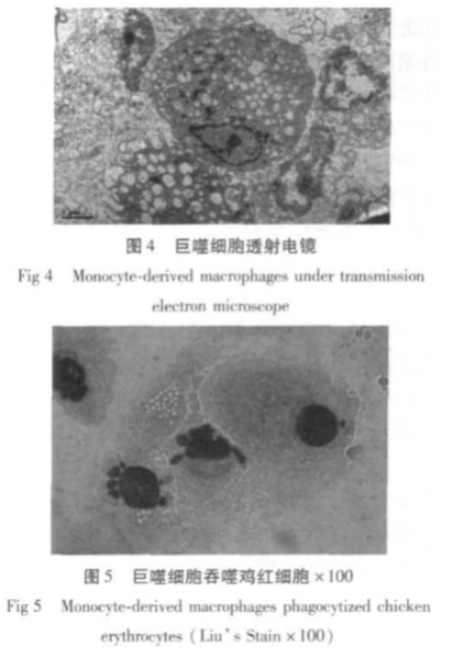
1. Transmission Electron Microscopy (TEM)
2. Gomori Acid Phosphatase (ACP) Staining: Calculate the percentage of ACP-positive macrophages.
3. Phagocytosis Assay:
Incubate macrophage suspension with 1% chicken red blood cells for 2 hours at 37°C.
Fix cell smears with methanol, stain with Giemsa, and observe under a microscope.
Calculate phagocytosis rate and index:
Phagocytosis rate = (Number of macrophages phagocytosing red blood cells / 100) × 100%
Phagocytosis index = Total number of red blood cells phagocytosed by 100 macrophages / 100
4. Flow Cytometry:
Detect CD14 expression using CD14-FITC and IgG2-FITC.
For M1 and M2 macrophages, detect markers such as TNF-α, CD86, CD11c, and CD206.
Dendritic Cell
Materials and Instruments
Reagents: 70% ethanol, RPMI-1640 medium, 1× PBS, ammonium chloride, GM-CSF, IL-4, FITC-anti-mouse CD11c, PE-anti-mouse CD40, FITC-anti-mouse CD80, PE-anti-mouse CD86, FITC-anti-mouse MHC II, isotype control antibodies (FITC-Rat IgG2b, PE-Rat IgG2a), IL-12p70, IFN-γ, IL-4, etc.
Instruments and consumables: Syringes, mouse dissection table, centrifuge, centrifuge tubes, flow cytometer, 200-mesh nylon mesh, cell culture dishes, cell counter, etc.
Procedure
1. Euthanize 6 C57BL/6 mice by cervical dislocation and disinfect with 70% ethanol for 5 minutes. Aseptically extract femurs and tibias, remove surrounding muscle tissue, wash twice with PBS, rinse once with incomplete 1640 medium, and transfer to a sterile dish on ice.
2. Fix the long bones with forceps, cut off both ends, and flush out the bone marrow using a syringe filled with incomplete 1640 medium. Repeat until the marrow turns white.
3. Collect the flushed medium, filter through a 200-mesh nylon mesh to remove muscle tissue and bone fragments. Centrifuge the filtrate at 1000 rpm for 5 minutes and discard the supernatant.
4. Add 10 ml of ammonium chloride red blood cell lysis buffer, incubate at room temperature for 2-3 minutes, and centrifuge at 1100 rpm for 5 minutes. Discard the supernatant.
5. Wash twice with incomplete 1640 medium, resuspend in 5 ml of complete medium, and plate in a culture dish.
6. Incubate at 37°C with 5% CO2 for 3 hours. Collect floating and semi-adherent cells, centrifuge at 1000 rpm for 5 minutes, and discard the supernatant.
7. Resuspend cells in RPMI-1640 medium with 10% FBS, count, and adjust density to 6 × 10^5 cells/ml. Plate in 24-well plates and add mouse GM-CSF (20 ng/ml) and IL-4 (10 ng/ml).
8. Incubate at 37°C with 5% CO2 (Day 0).
9. On Day 2, gently shake the plate, remove 1/2 to 2/3 of the old medium (to remove granulocytes), and replenish with fresh medium and cytokines every two days.
10. On Day 7, gently pipette to collect floating and loosely adherent cells, which are immature DCs.
11. Centrifuge at 1000 rpm for 10 minutes, discard the supernatant, resuspend in medium, and adjust density to 1 × 10^6 cells/ml. Plate in 24-well plates and add LPS (1 µg/ml), TNF-α (250 U/ml), or CD40L (1 µg/ml). After 48 hours, collect floating and loosely adherent cells as mature DCs. Collect and freeze supernatants at -70°C for IL-12p70 detection.
Cell Detection
1. Light and Electron Microscopy
2. Surface Marker Detection:
Use FITC-anti-mouse CD11c, PE-anti-mouse CD40, FITC-anti-mouse CD80, PE-anti-mouse CD86, FITC-anti-mouse MHC II, and isotype controls (FITC-Rat IgG2b, PE-Rat IgG2a). Incubate at 4°C for 30 minutes, wash with staining buffer, and analyze by flow cytometry.
3.Mixed Lymphocyte Reaction (MLR):Co-culture mature or immature DCs with CD4+ T cells for 72 hours. Add 3H-TdR 16 hours before termination. Collect cells onto fiber membranes, add scintillation fluid, and measure cpm values.
4.ELISA for Cytokines:
Coat 96-well plates with capture antibodies. Use IL-12p70, IFN-γ, and IL-4 standards as controls. Add culture supernatants to test wells and measure cytokine concentrations.
Natural Killer (NK) Cell Culture
Materials and Instruments
Reagents: Recombinant human IL-2, recombinant human IL-15, RPMI-1640 medium, CD3-FITC, CD45-PerCP, CD56-PE, Ficoll, 1× PBS.
Instruments: OLYMPUS microscope, flow cytometer, CO2 incubator, biosafety cabinet, centrifuge, ultrapure water system, water bath, etc.
Procedure
1. In a biosafety cabinet, dilute 15 ml of PBMC sample 1:1 with saline.
2. Add 30 ml of PBMC sample to a tube and mix with Ficoll at a 2:1 ratio.
3. Centrifuge at 2000 rpm for 20 minutes.
4. Carefully collect the plasma layer and the white cell layer into separate tubes.
5. Incubate the plasma tube at 56°C for 30 minutes, then centrifuge at 3000 rpm for 15 minutes. Collect the supernatant.
6. Wash the white cell layer with 1× PBS, centrifuge at 2100 rpm for 6 minutes, discard the supernatant, and repeat.
7. Count cells and seed at 1-2 × 10^6 cells/ml in CD16 antibody-coated T75 flasks. Add 10% heat-inactivated autologous plasma, 1000 U/ml IL-2, and 50 ng/ml IL-15.
8. Incubate at 37°C with 5% CO2.
9. Monitor cell growth on Days 2, 3, and 5, replenishing medium, plasma, and cytokines as needed.
10. On Day 7, count cells. If density exceeds 2 × 10^8 cells/ml, transfer to a culture bag.
11. Collect cells for analysis between Days 8-10.
Cell Detection
1.Trypan Blue Staining: Ensure >90% viability.
2.Flow Cytometry:
Collect cells on Days 7 and 10, adjust density to 1 × 10^7 cells/ml, and stain with CD3-FITC, CD45-PerCP, and CD56-PE. Include isotype controls. Incubate at 4°C for 30 minutes, wash with PBS, and analyze by flow cytometry.
3.Cytotoxicity Assay:
Collect NK cells on Day 10, label with CFSE, and co-culture with K562 cells at ratios of 5:1, 10:1, and 20:1. Include NK and K562 cell controls. After 4 hours, measure K562 cell death using 7-AAD staining.
UA BIOSCIENCE offers high-quality cytokines with validated activity, suitable for the culture of various immune cells, stem cells, and organoids. Rest assured, our products are of exceptional quality! (Ensure cytokine selection matches the sample source!)
This article is reviewed and published by the technical expert team of UA
Disclaimer: This article partially utilizes artificial intelligence assistance in its creation. If any content involves copyright or intellectual property issues, please let us know and we promise to verify and remove it as soon as possible.
Purchase recombinant protein, choose Nanjing UA-Bio
UA protein focuses on providing various protein reagents, raw materials, and services required for drug research and development, cell therapy, gene therapy, and basic scientific research, including drug target proteins, immune checkpoint proteins, cytokines, tool enzymes, customized protein expression, and full-length transmembrane protein development. Youai is committed to providing customers with high-quality products and professional services, and building a High-tech Biological Enterprise with International Competitiveness.

1.The Impact of Cytokine Pretreatment on the Activity of NK Cells in In Vitro Culture.
2.Webpage Compilation






 Business license (three certificates in one)