




Revitalizing Insights: New Discoveries in the Biology and Clinical Applications of IL-18


IL-18 is a member of the IL-1 family of cytokines, renowned for its potent ability to induce IFNγ production. However, both fundamental research and recent clinical observations suggest a more complex scenario. The unique biological characteristics of IL-18 at the levels of transcription, activation, secretion, neutralization, receptor distribution, and signaling contribute to explaining its pleiotropic roles in mucosal and systemic inflammation.
- Recent Advances
- Product Information
- Reference
Introduction
Several recent discoveries have reignited interest in the pathogenic potential and possible clinical applications of IL-18. IL-18 is a member of the IL-1 family of cytokines, renowned for its potent ability to induce IFNγ production. However, both fundamental research and recent clinical observations suggest a more complex scenario. The unique biological characteristics of IL-18 at the levels of transcription, activation, secretion, neutralization, receptor distribution, and signaling contribute to explaining its pleiotropic roles in mucosal and systemic inflammation. Blood biomarker studies have revealed a cytokine whose significant elevation is associated with detectable "free IL-18," defining a group of autoinflammatory diseases where IL-18 dysregulation may be a primary driving feature, termed "IL-18opathies." This impressive specificity may accelerate diagnosis and identify patients suitable for therapeutic IL-18 blockade. Pathologically, human and animal studies have found that CD8+ T cells are preferentially activated over other IL-18-responsive lymphocytes. Therapeutic strategies utilizing IL-18 agonists that exploit production sites or subvert endogenous IL-18 inhibition show promise in enhancing cancer immune responses. Thus, the unique aspects of IL-18 biology are finally beginning to have clinical impacts in the precise diagnosis, disease monitoring, and targeted treatment of inflammatory and malignant diseases.
About IL-18
Between transcription and release, the processes of translation to secretion for IL-1 family members IL-1β and IL-18 are closely matched. Pro-IL-1β and Pro-IL-18 are inactive precursors that enter the cytoplasm. Both require proteolytic cleavage to become active cytokines and rely on membrane pores to exit their cells of origin. However, differences in pre-translational and post-release events have biological and clinical significance.
IL-18 is often considered a macrophage-derived cytokine, but current data indicate that IL-18 production varies significantly depending on the tissue and inflammatory signals received by macrophages. Moreover, nearly all barrier epithelia also contain substantial amounts of IL-18, sometimes acting as inducible effectors and other times as pre-formed alarms. The implications of this pleiotropy are only beginning to be elucidated.
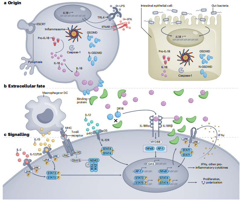
Figure 1: Origin, Extracellular Context, and Signaling of IL-18
a. Whether constitutively produced (epithelial cells and some tissue macrophages) or induced by complex stimuli (e.g., lipopolysaccharide (LPS), interferon (IFN), or intestinal dysbiosis), pro-IL-18 is typically cleaved by caspase-1 (or caspase-8, granzyme-B, proteinase-3, or chymase) into its active form, allowing it to exit the cell through gasdermin D (GSDMD) pores in an inflammasome-dependent manner. The ESCRT pathway can prevent pyroptosis caused by GSDMD pores.
b. Once released, IL-18 is usually bound and neutralized by IL-18 binding protein (IL-18BP), which is itself produced by most cell types in response to IFNγ. Non-bound or "decoy-resistant" IL-18 (DR-18) signals through the IL-18 receptor heterodimer (IL-18R), often acting synergistically with other signals.
c. IL-18R signals through the traditional IL-1 family/MyD88-dependent pathway. IL-18 can drive NF-κB and/or MAPK signaling, promoting proliferation, pro-inflammatory cytokine expression, and/or other effector programs (e.g., cytotoxicity). The diverse effects of IL-18 depend on the type and state of the cells it acts upon, as well as the presence of other signals such as TCR, IL-2, myeloid-derived IL-15, IL-12, and/or IFNγ (forming a positive feedback loop).
Applications of IL-18
IL-18 as a Biomarker
Clinicians routinely interpret levels of acute-phase reactants and inflammatory biomarkers. While most of these tests are valuable for monitoring disease activity, they lack specificity. Measurement of total IL-18 is rapidly becoming essential for diagnosing a subset of rheumatic and/or autoinflammatory diseases, particularly in pediatric rheumatology.
IL-18 and Disease
In many cases, profound and chronic elevations in total IL-18 (accompanied by detectable free IL-18) delineate a spectrum of primarily autoinflammatory diseases appropriately termed "IL-18opathies." Most of these diseases involve an increased susceptibility to macrophage activation syndrome (MAS), a secondary form of hemophagocytic lymphohistiocytosis (HLH) associated with rheumatic diseases. HLH and MAS are cytokine storm syndromes characterized by fever, hyperferritinemia (often >1,000 ng/ml), peripheral cytopenia, hepatosplenomegaly, hepatitis, disseminated intravascular coagulation, central nervous system involvement, and frequently, organ failure and death. Severe genetic defects in granule-mediated cytotoxicity are often described as the prototypical cause of HLH, although such defects account for only a small fraction of all HLH cases.

Figure 2: Pathological Manifestations of IL-18opathies Associated with IL-18 Sources
a. Although fever is less prominent in PSTPIP1-associated diseases, signs of systemic inflammation are present in all IL-18opathies.
b. Pericarditis occurs in some patients with systemic juvenile idiopathic arthritis/adult-onset Still's disease (sJIA/AOSD).
c. Colitis and inflammatory bowel disease (IBD) occur in patients with severe NLRC4 hyperactivity or XIAP deficiency. Colonic and small intestinal epithelial cells produce substantial amounts of pro-IL-18 under steady-state conditions.
d. Hepatitis, hepatosplenomegaly, and hemophagocytosis can occur in patients with macrophage activation syndrome/hemophagocytic lymphohistiocytosis (MAS/HLH) of any cause. Hepatic Kupffer cells and splenic red pulp macrophages can produce significant amounts of pro-IL-18.
e. Cutaneous manifestations of IL-18opathies may include various transient, urticarial, or nodular rashes in sJIA/AOSD and monogenic MAS patients. PSTPIP1-associated diseases present with cystic acne and pyoderma gangrenosum, but MAS has not been reported in these patients. Keratinocytes and Langerhans cells produce substantial amounts of pro-IL-18 under steady-state conditions.
f. Pulmonary alveolar proteinosis (PAP) with endogenous lipoid pneumonia (ELP) has been observed in sJIA-associated lung disease. Both respiratory epithelial cells and alveolar macrophages produce significant amounts of IL-18 under steady-state conditions.
g. Tumors secrete large amounts of IL-18BP, which prevents IL-18 from promoting anti-tumor immunity but can be overcome by "decoy-resistant" IL-18 (DR-18) or IL-18-producing CAR-T cells.
h. Septic arthritis occurs in many patients with PSTPIP1-associated diseases. Although arthritis is a diagnostic criterion for sJIA/AOSD, its incidence is lower in sJIA/AOSD patients with higher IL-18 levels.
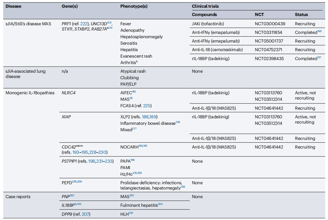
IL-18-Related Diseases and Ongoing Clinical Studies
(Currently, 461 clinical studies related to IL-18 are registered in NCBI.)
Future Perspectives
Exploring the mechanisms and clinical applications of IL-18 in inflammatory diseases can provide critical insights for the diagnosis and treatment of related conditions. The measurement of IL-18 concentrations and modulation of IL-18 signaling pathways may emerge as novel therapeutic targets in the future.
In summary, IL-18 plays a significant role in the pathogenesis and treatment of inflammatory diseases. A deeper understanding of its biological functions and clinical implications will advance the diagnosis and management of related conditions.
This article is reviewed and published by the technical expert team of UA
Disclaimer: This article partially utilizes artificial intelligence assistance in its creation. If any content involves copyright or intellectual property issues, please let us know and we promise to verify and remove it as soon as possible.
Purchase recombinant protein, choose Nanjing UA-Bio
UA protein focuses on providing various protein reagents, raw materials, and services required for drug research and development, cell therapy, gene therapy, and basic scientific research, including drug target proteins, immune checkpoint proteins, cytokines, tool enzymes, customized protein expression, and full-length transmembrane protein development. Youai is committed to providing customers with high-quality products and professional services, and building a High-tech Biological Enterprise with International Competitiveness.

1.Emily L. et al.Biological and clinical roles of IL-18 in inflammatory diseases.Nature Reviews Rheumatology20,pages33-47(2024).
2.Bossaller, L. et al. Cutting edge: FAS (CD95) mediates noncanonical IL-1β and IL-18 maturation via caspase-8 in an RIP3-independent manner. J. Immunol. 189, 5508–5512 (2012).
3. Omoto, Y. et al. Granzyme B is a novel interleukin-18 converting enzyme. J. Dermatol. Sci.59, 129–135 (2010).
4. Sugawara, S. et al. Neutrophil proteinase 3-mediated induction of bioactive IL-18 secretion by human oral epithelial cells. J. Immunol. 167, 6568–6575 (2001).
35. Omoto, Y. et al. Human mast cell chymase cleaves pro-IL-18 and generates a novel and biologically active IL-18 fragment. J. Immunol. 177, 8315–8319 (2006).
5. Aguilar, C. et al. Characterization of Crohn disease in X-linked inhibitor of apoptosis-deficient male patients and female symptomatic carriers. J. Allergy Clin. Immunol. 134, 1131–1141.e9 (2014).
6.Canna,S.W. et al. Brief report: alternative activation of laser-captured murine hemophagocytes. Arthritis Rheumatol. 66, 1666–1671 (2014).
7. Billiau, A. D., Roskams, T., Van Damme-Lombaerts, R., Matthys, P. & Wouters, C. Macrophage activation syndrome: characteristic findings on liver biopsy illustrating the key role of activated, IFN-γ-producing lymphocytes and IL-6- and TNF-α-producing macrophages. Blood 105, 1648–1651 (2005).






 Business license (three certificates in one)