




Molecular characteristics and signal transduction mechanisms of tumor necrosis factor receptor 1


Tumor necrosis factor receptor 1 (TNFR1, also known as TNFRSF1A, CD120a, or p55) is an important member of the tumor necrosis factor receptor superfamily.
- Recent Advances
- Product Information
1. What are the molecular structure and expression characteristics of TNFR1?
Tumor necrosis factor receptor 1 (TNFR1, also known as TNFRSF1A, CD120a, or p55) is a key member of the tumor necrosis factor receptor superfamily. As a 55 kDa type I transmembrane protein, it is widely expressed on the surface of various cells in the body, particularly showing high expression in immune cells. Its molecular structure consists of three critical regions: the extracellular region contains four cysteine-rich domains (CRD1-CRD4), with CRD2 and CRD3 forming the binding site for TNF-α; the transmembrane region consists of 22 amino acids; and the intracellular region includes important functional modules such as the death domain (DD), neutral sphingomyelinase domain (NSD), and TNFR1 internalization domain (TRID). The expression of TNFR1 is finely regulated by various elements in its gene promoter region, maintaining constitutive expression at basal levels while being inducibly upregulated via pathways like NF-κB in response to inflammatory stimuli, ensuring timely responses during immune reactions.
2. What is the binding specificity between TNFR1 and TNF-α?
TNF-α exists in two distinct forms: the 26 kDa transmembrane form (tmTNF) and the 17 kDa soluble form (solTNF), which achieve functional differentiation through binding to different receptors. TNFR1 primarily binds to soluble TNF-α. When the solTNF trimer specifically interacts with the CRD2 and CRD3 domains of TNFR1's extracellular region, it induces the receptor to form a trimeric complex. This binding relies on the pre-ligand binding assembly domain (PLAD)-driven pre-assembly of receptors, ensuring efficient signal initiation. In contrast, transmembrane TNF-α preferentially binds to TNFR2, mediating tissue repair and immunomodulatory signals. This receptor-ligand pairing specificity makes TNFR1 the primary receptor mediating TNF-α's pro-inflammatory and pro-apoptotic effects.
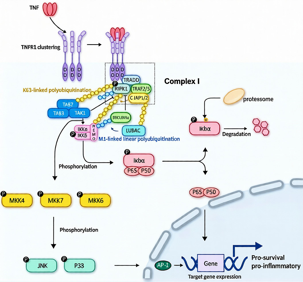
3. How does the TNFR1-mediated signal transduction pathway operate?
TNFR1 signal transduction achieves functional differentiation through the dynamic assembly of different intracellular complexes. Upon TNF-α binding, complex I (also known as the membrane-associated complex) forms first, with TRADD (TNFR1-associated death domain protein) serving as the core scaffold, recruiting signaling molecules such as RIPK1, TRAF2/5, and cIAP1/2 to form an E3 ubiquitin ligase complex. During this process, RIPK1 undergoes K63-linked ubiquitination, subsequently recruiting and activating the TAK1/TAB complex and the IKK complex (comprising IKKα, IKKβ, and NEMO subunits). Activated TAK1 initiates the MAPK signaling cascade (including JNK and p38 pathways), while the IKK complex phosphorylates IκB proteins, leading to their degradation and the release of NF-κB transcription factors into the nucleus to initiate the expression of pro-inflammatory and pro-survival genes.
Under specific conditions, TNFR1 signaling can shift to the assembly of complex II (also known as the cytoplasmic complex), where TRADD dissociates from the receptor and forms an apoptotic signaling complex with FADD and caspase-8, initiating the apoptotic program. Additionally, TNFR1 can induce ceramide production via the neutral sphingomyelinase domain, triggering mitochondrial dysfunction and leading to another form of cell death. The balance of these signaling pathways determines the cell's ultimate fate—survival and proliferation or programmed death.
4. What are the key applications of TNF-α/TNFR1 detection kits?
Accurate detection of TNF-α and its receptors holds significant importance in clinical diagnosis and basic research. The primary applications of TNF-α/TNFR1 detection kits include:
1. Disease Diagnosis and Stratification: By measuring soluble TNFR1 levels in serum, plasma, or synovial fluid samples, these kits can assist in evaluating disease activity and severity in autoimmune diseases such as rheumatoid arthritis and inflammatory bowel disease, providing objective criteria for clinical classification.
2. Treatment Monitoring and Prognostic Assessment: In patients receiving anti-TNF-α biologic therapies, dynamic monitoring of TNF-α and soluble TNFR1 level changes can assess treatment efficacy, predict therapeutic responses, and guide treatment adjustments. Studies show that changes in soluble TNFR1 levels before and after treatment are closely correlated with clinical outcomes.
3. Drug Development Support: In the development of novel drugs targeting the TNFR1 signaling pathway, highly sensitive and specific detection kits can be used to screen candidate compounds, evaluate pharmacodynamic characteristics, and elucidate mechanisms of action, accelerating the drug development process.
4. Basic Mechanism Research: When studying TNF-α/TNFR1 signaling mechanisms or exploring the pathway's role in disease pathogenesis, reliable quantitative detection tools are essential for in-depth research, providing critical data to elucidate pathological mechanisms.
5. Which manufacturers provide TNF-α/TNFR1 detection kits?
Nanjing U-Protein Biotechnology has independently developed the "TR-FRET Human TNF-α/TNFR1 Binding Kit" (Catalog No.: UA086007), a high-sensitivity, homogeneous detection platform based on advanced time-resolved fluorescence resonance energy transfer (TR-FRET) technology. This kit focuses on the precise quantification of the in vitro binding activity between tumor necrosis factor-α (TNF-α) and its key receptor TNFR1, providing an efficient, stable, and standardized solution for autoimmune diseases, chronic inflammation, TNF signaling pathway research, and related antibody/small-molecule drug development, with compatibility for high-throughput applications.
| Core Product Advantages |
|---|
| High Sensitivity and Superior Signal-to-Noise Ratio: Leveraging TR-FRET technology, the kit achieves time-resolved and dual-wavelength detection, effectively eliminating sample background fluorescence and compound interference, significantly enhancing detection sensitivity and signal-to-noise ratio, particularly suitable for analyzing weak binding interactions and complex sample systems. |
| Native Conformation and High Bioactivity: The kit provides rigorously validated high-purity, high-activity human TNF-α and TNFR1 proteins, both maintaining intact functional conformations and natural binding properties, ensuring binding experiments accurately simulate physiological signal initiation events. |
| Homogeneous Detection and Operational Convenience: Adopting a "mix-incubate-read" homogeneous detection mode, the kit eliminates the need for washing or separation steps, simplifying the workflow and enhancing compatibility with automated workstations, thereby significantly improving experimental efficiency and data reproducibility. |
| Excellent Stability and Batch-to-Batch Consistency: Through advanced recombinant expression systems and stringent quality control, the protein products exhibit high purity, superior long-term stability, and exceptional batch-to-batch consistency, providing reliable support for long-term and continuous drug screening and mechanistic studies. |
| Comprehensive Solutions and Professional Support: We provide detailed and optimized experimental protocols, standard curve examples, and data analysis guidelines, along with professional technical support and customized services tailored to specific research needs (e.g., competitive inhibitor evaluation, antibody epitope analysis). |
Nanjing U-Protein Biotechnology is committed to providing cutting-edge and reliable research tools and solutions for inflammation, immunology, and innovative drug development. For detailed technical information, validation data, or application support regarding the TR-FRET Human TNF-α/TNFR1 Binding Kit (Catalog No.: UA086007), please feel free to contact us.
This article is reviewed and published by the technical expert team of UA
Disclaimer: This article partially utilizes artificial intelligence assistance in its creation. If any content involves copyright or intellectual property issues, please let us know and we promise to verify and remove it as soon as possible.
Purchase recombinant protein, choose 
UA protein focuses on providing various protein reagents, raw materials, and services required for drug research and development, cell therapy, gene therapy, and basic scientific research, including drug target proteins, immune checkpoint proteins, cytokines, tool enzymes, customized protein expression, and full-length transmembrane protein development. Nanjing Univ is committed to providing customers with high-quality products and professional services, and building a High-tech Biological Enterprise with International Competitiveness.







 Business license (three certificates in one)

