




DNA Repair Enzymes PARPs: A Comprehensive Analysis from Molecular Mechanisms to Clinical Applications


In the intricate world of cells, DNA repair enzymes known as PARPs (Poly ADP-Ribose Polymerases) play a pivotal role. These enzymes are not only critical participants in DNA damage repair but also exert significant functions in processes such as apoptosis, gene transcription, and DNA synthesis. This article delves into the functions, mechanisms of action, and biomedical applications of DNA repair enzyme PARPs.
- Recent Advances
- Product Information
- Reference
Introduction
In the intricate world of cells, DNA repair enzymes known as PARPs (Poly ADP-Ribose Polymerases) play a pivotal role. These enzymes are not only critical participants in DNA damage repair but also exert significant functions in processes such as apoptosis, gene transcription, and DNA synthesis. This article delves into the functions, mechanisms of action, and biomedical applications of DNA repair enzyme PARPs.
Overview of the PARP Family
The PARP family consists of 17 members, divided into five subgroups based on domain structure and function: DNA damage-dependent PARPs (PARP1, PARP2, and PARP3), tankyrase 1/PARP5 and tankyrase 2/PARP5b, CCCH-type PARPs (PARP7, PARP12, and PARP13), macroPARPs (BAL1/PARP9, BAL2/PARP14, and BAL3/PARP15), and other PARPs (PARP4, PARP6, PARP8, PARP10, PARP11, and PARP16). The C-terminal catalytic domain is conserved across all members and includes additional zinc fingers, BRCA C-terminal-like (BRCT) motifs, ankyrin repeats, macrodomains, and WWE domains.
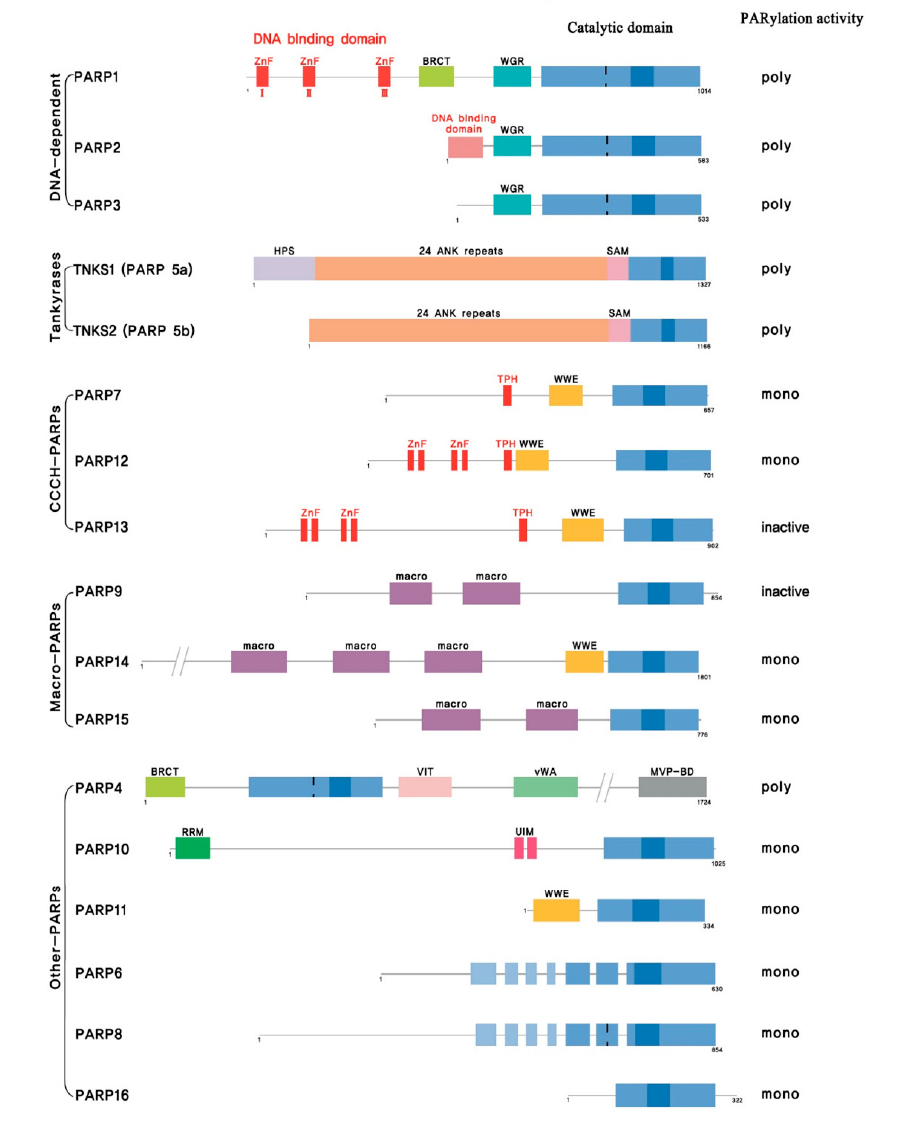
Figure 1: PARP Family Source: cancers
Mechanism of PARP1
PARylation is a reaction catalyzed by ADP-ribosyltransferases, which utilize NAD+ to transfer ADP-ribose residues to target substrates. PARPs, particularly PARP1, PARP2, PARP3, PARP5a, and PARP5b, rapidly recognize various types of DNA damage, including single-strand breaks (SSBs), and are recruited to damage sites to facilitate the recruitment of DNA damage response (DDR) molecules. PARP1, the most prominent member of the PARP family, accounts for over 90% of PARP functions in cells and is a key factor in DNA damage repair.
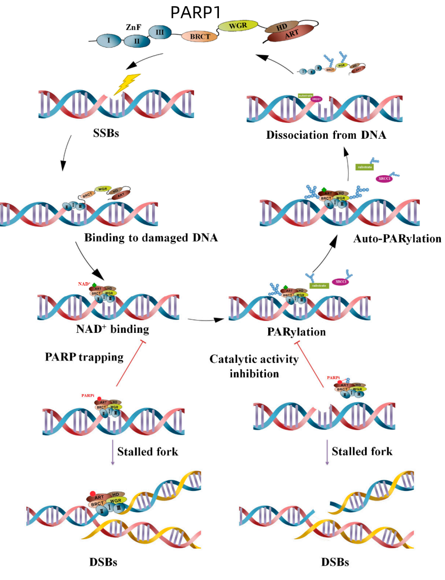
Figure 2: Mechanism of PARylation and PARP Inhibition in DNA Damage Response Source: cancers
When a single-strand break occurs, PARP rapidly binds to the damage site via its zinc finger domain. Through its ART catalytic domain, PARP uses NAD+ as a substrate to catalyze PARylation between itself and target proteins such as XRCC1 and DNA ligase III, recruiting DNA repair proteins to the damage site. Spontaneous PARylation of PARP reduces its affinity for DNA, causing it to dissociate and allowing repair proteins to bind. PARP inhibitors, however, bind to the catalytic pocket instead of NAD+, trapping PARP on DNA. This leads to replication fork stalling, conversion to double-strand breaks (DSBs), and ultimately cell death. Alternatively, PARP inhibitors can block the recruitment of repair proteins by inhibiting PARP's enzymatic activity.
Applications of PARPs in Biomedicine
Due to their critical roles in DNA repair and apoptosis, PARPs have become important targets in biomedical research. Several PARP inhibitors have been developed for the treatment of diseases such as cancer. These inhibitors block PARP activity, thereby inhibiting cancer cell growth and proliferation while promoting apoptosis and enhancing therapeutic efficacy.
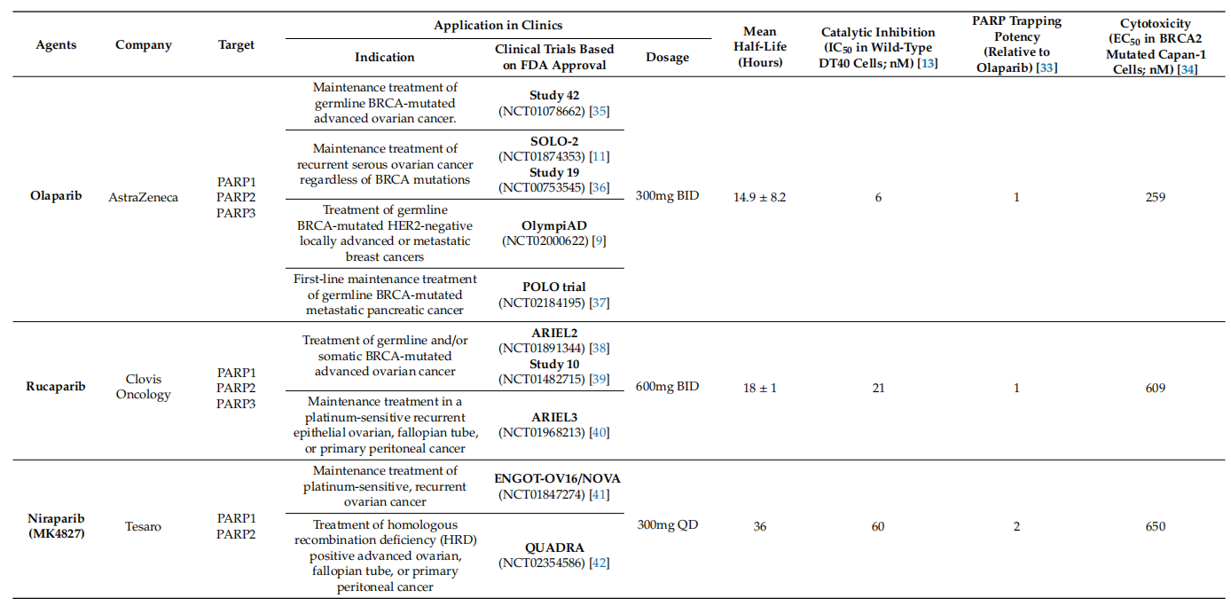
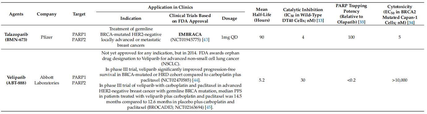
Table 1: Comparison of Clinically Available PARP Inhibitors
PARP inhibitors have shown efficacy not only in BRCA-mutated tumors but also in platinum-sensitive tumors caused by homologous recombination deficiency (HRD), accelerating their clinical application. However, the effects of these inhibitors cannot be fully explained by the inhibition of PARP catalytic activity alone. Although all four PARP inhibitors inhibit the catalytic activity of PARP1 and PARP2, their cytotoxicities differ, as shown in Table 1. The cytotoxicity induced by PARP inhibitors far exceeds that induced by PARP gene knockout, suggesting that their anti-tumor effects involve mechanisms beyond catalytic inhibition. This difference can be conceptualized as PARP trapping: the ability of PARP inhibitors to stabilize PARP-DNA complexes, increasing their binding stability. As shown in Table 1, the cytotoxicity of each PARP inhibitor correlates with its PARP trapping activity. For example, talazoparib exhibits the strongest PARP trapping activity and the highest cytotoxicity. Therefore, PARP trapping should be considered a mechanism of action for PARP inhibitors in clinical applications. These differences in PARP trapping capabilities may influence the outcomes of combination and monotherapy.
PARPs have diverse functions, ranging from transcriptional regulation to post-translational modifications that activate and localize proteins. In DNA damage response, PARPs regulate the activation of themselves or their target proteins through PARylation. PARP inhibitors block this catalytic activity, preventing the activation of normal repair pathways. These inhibitors have shown significant anti-tumor effects in HRD tumors, such as those with BRCA mutations. To date, four PARP inhibitors have been FDA-approved and used in clinical practice. However, these compounds differ in their ability to trap PARP-DNA complexes, despite sharing the common feature of inhibiting PARP catalytic activity. PARP trapping improves cytotoxicity by causing replication fork collapse, leading to DSBs. When combined with alkylating agents, PARP inhibitors exhibit synergistic effects. Conversely, inhibiting PARP catalytic activity shows synergy with topoisomerase I inhibitors. Thus, the synergistic effects of combination therapy with PARP inhibitors can vary depending on the specific mechanism of action of each inhibitor. Understanding the unique properties of each PARP inhibitor and strategically selecting synergistic partners are critical considerations for maximizing anti-tumor efficacy.
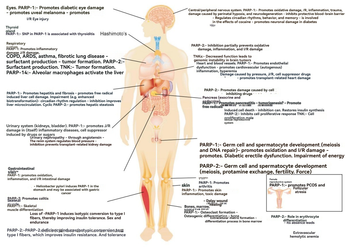
Figure 3: Role of PARPs in Human Organs Source: Cell Press
ARDS, acute respiratory distress syndrome; COPD, chronic obstructive pulmonary disease; I/R, ischemia-reperfusion; PCOS, polycystic ovary syndrome.
These diseases affect all major organ systems, including ischemia-reperfusion injury, inflammatory diseases, burns, Parkinson's disease, Huntington's disease, Alzheimer's disease, and toxic injuries (e.g., cytotoxicity from inhibitors, cigarette smoke, UV, and gamma radiation).
Conclusion
PARPs hold broad application prospects in cancer therapy, antiviral innate immunity, gene editing, genetic disease treatment, and other biomedical fields. With further research into the functions and mechanisms of PARPs, we anticipate the development of more PARP-targeted drugs and therapies, contributing significantly to human health.
This article is reviewed and published by the technical expert team of UA
Disclaimer: This article partially utilizes artificial intelligence assistance in its creation. If any content involves copyright or intellectual property issues, please let us know and we promise to verify and remove it as soon as possible.
Purchase recombinant protein, choose Nanjing UA-Bio
UA protein focuses on providing various protein reagents, raw materials, and services required for drug research and development, cell therapy, gene therapy, and basic scientific research, including drug target proteins, immune checkpoint proteins, cytokines, tool enzymes, customized protein expression, and full-length transmembrane protein development. Youai is committed to providing customers with high-quality products and professional services, and building a High-tech Biological Enterprise with International Competitiveness.

1. Ahrum Min.et al. PARP Inhibitors as Therapeutics: Beyond Modulation of PARylation.Cancers.2020.
2. O’Neil,N.J.et al.Syntheticle thalityand cancer.Nat.Rev.Genet.2017,18,613–623.
3. Beijersbergen,R.et al.Synthetic Lethalityin Cancer Therapeutics.Annu.Rev.Cancer Boil.2017,1,141–161.
4. Murai, J.et al. Trapping of PARP1 and PARP2 by Clinical PARP Inhibitors. Cancer Res. 2012, 72, 5588–5599.
5. Murai, J.et al. Stereospecific PARP trapping by BMN 673 and comparison with olaparib and rucaparib.Mol. Cancer Ther. 2014, 13, 433–443.
6. Shen, Y. et al. Trapping Poly(ADP-Ribose) Polymerase. J. Pharmacol. Exp. Ther.2015, 353, 446–457.
7. Kaufman, B. et al. Olaparib Monotherapy in Patients With Advanced Cancer and a Germline BRCA1/2 Mutation. J. Clin. Oncol. 2015, 33, 244–250.
8. Peter Bai. et al.Biology of Poly(ADP-Ribose) Polymerases:The Factotums of Cell Maintenance.CellPress.2015.






 Business license (three certificates in one)