




The mechanism of TNF-α/TNFR1 signaling pathway and its application in the development of detection kits


The tumor necrosis factor α (TNF-α) and its receptor TNFR1 signaling pathway constitute a core molecular system that regulates inflammation, cell survival, and programmed cell death.
- Recent Advances
- Product Information
Abstract
The signaling pathway formed by tumor necrosis factor α (TNF-α) and its receptor TNFR1 constitutes the core molecular system regulating inflammation, cell survival, and programmed cell death. TNF-α exists in two forms: membrane-bound (tmTNF) and soluble (sTNF), preferentially activating TNFR2 and TNFR1 respectively, mediating dual biological effects of protective repair and pro-inflammatory damage. As a type I transmembrane protein, TNFR1 binds ligands through its extracellular CRD domains and dynamically assembles signaling complexes via intracellular death domains and other modules, precisely regulating NF-κB-mediated pro-inflammatory survival signals and death pathways such as apoptosis, necroptosis, and pyroptosis. Based on in-depth understanding of this pathway, TNF-α/TNFR1 detection kits have become important tools for studying inflammatory mechanisms, disease diagnosis, and drug development.
I. Overview of the TNF-α/TNFR1 System
1. Molecular Structure and Expression Characteristics of TNFR1
TNFR1 (CD120a, TNFRSF1A) is a 55 kDa type I transmembrane protein. Its extracellular region contains four cysteine-rich domains (CRD1-4), with CRD2 and CRD3 being key regions for TNF-α binding. The intracellular region includes functional modules such as the death domain, neutral sphingomyelinase domain, and internalization domain, responsible for signal transduction and functional differentiation. TNFR1 is widely expressed in most human tissues, particularly at high levels in immune cells, and its expression is dynamically regulated by transcription factors such as NF-κB and AP-1.
2. Two Forms of TNF-α and Receptor Selectivity
- Membrane-bound TNF (tmTNF): Anchored to the cell membrane as a precursor, it primarily binds to TNFR2 to transmit protective signals such as tissue repair and immune regulation.
- Soluble TNF (sTNF): Released from tmTNF via enzymatic cleavage, it preferentially activates TNFR1, driving pro-inflammatory responses and cell death signals.
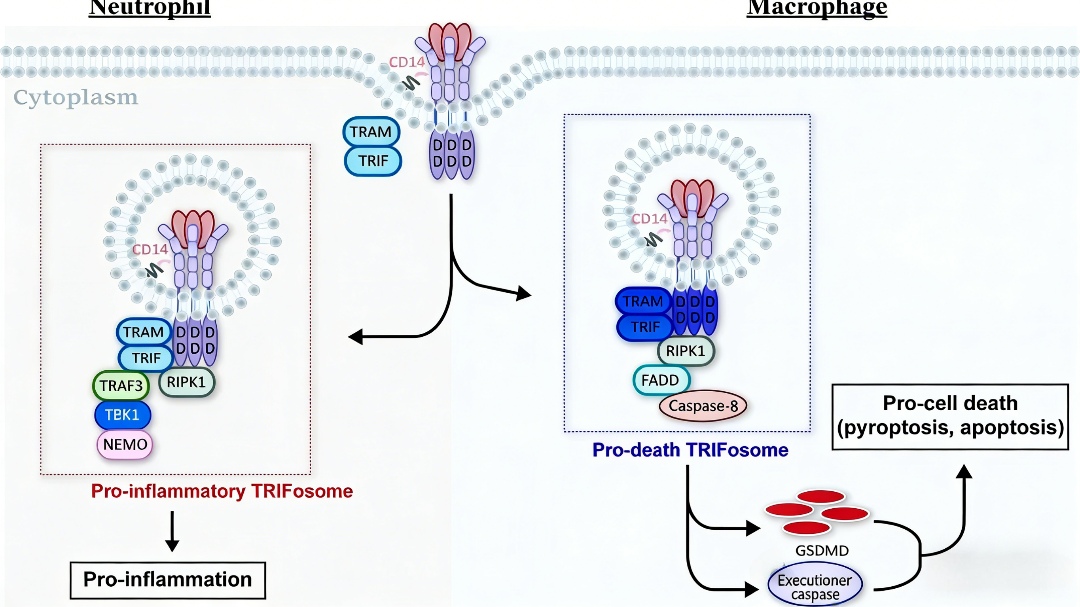
II. Core Mechanisms of TNFR1 Signal Transduction
1. Complex I: Pro-inflammatory and Pro-survival Signaling Pathway
TNF-α induces TNFR1 trimerization, recruiting proteins such as TRADD, RIPK1, TRAF2/5, and cIAP1/2 to form the membrane-associated Complex I. This complex activates TAK1 and the IKK complex through K63 ubiquitination, subsequently activating MAPK and NF-κB pathways, promoting inflammatory factor expression and cell survival.
2. Complex II: Initiation and Differentiation of Cell Death Signals
Under specific conditions (e.g., abnormal ubiquitination or caspase inhibition), RIPK1 forms cytoplasmic Complex II with FADD, caspase-8, etc., initiating different death programs:
- Apoptosis: Caspase-8 activates downstream effector caspases, leading to programmed apoptosis.
- Necroptosis: When caspase-8 is inhibited, RIPK1 and RIPK3 form the necrosome, phosphorylating MLKL and disrupting plasma membrane integrity.
- Pyroptosis: Primarily occurs in immune cells, activating caspase-8 and GSDMD via the TRIF-dependent pathway, inducing inflammatory cell death.
III. Development and Applications of TNF-α/TNFR1 Detection Kits
Based on the molecular foundation of the above signaling pathways, TNF-α/TNFR1 detection kits typically include the following key components and design principles:
| Functional Module | Detection Content and Methods |
|---|---|
| 1. Detection Targets and Forms | - TNF-α Detection: Quantifies sTNF and tmTNF levels, distinguishing their expression patterns in different pathological states. - TNFR1 Detection: Evaluates receptor expression levels, cleavage forms, and activation states. - Signal Complex Analysis: Detects Complex I/II-related proteins (e.g., RIPK1 ubiquitination, caspase activation). |
| 2. Technology Platforms and Advantages | - ELISA and Chemiluminescence: Suitable for high-throughput quantification of TNF-α/TNFR1 in serum, cell supernatants, and other samples. - Flow Cytometry: Enables single-cell level analysis of receptor expression, internalization, and co-localization of signaling molecules. - Immunohistochemistry/Fluorescence: Used for spatial distribution and expression level studies of TNFR1 in tissue sections. |
| 3. Applications in Research and Clinical Settings | - Inflammation and Autoimmune Disease Research: Assesses disease activity, treatment response, and biomarker screening. - Tumor Immunoanalysis: Detects the regulation of immune cell function by TNF-α signaling in the tumor microenvironment. - Drug Development and Efficacy Evaluation: Used for screening and mechanistic validation of antibodies and small-molecule inhibitors targeting the TNF-α/TNFR1 pathway. |
IV. Summary and Outlook
As a key hub of TNF-α signaling, TNFR1 precisely regulates the balance between inflammation, survival, and death by dynamically assembling membrane complexes and cytoplasmic signaling platforms. Detection kits targeting this pathway not only provide standardized tools for basic research but also demonstrate significant value in disease diagnosis, efficacy monitoring, and drug development. With deepening understanding of the signaling regulatory network and continuous optimization of detection technologies, the TNF-α/TNFR1 detection system will play a broader role in precision medicine and translational research.
V. Which Manufacturers Provide TNF-α/TNFR1 Detection Kits?
Nanjing UniProtein independently developed the TR-FRET Human TNF-α/TNFR1 Binding Kit (Catalog No.: UA086007), a high-performance detection platform based on advanced time-resolved fluorescence energy transfer (TR-FRET) technology, specifically designed for accurate quantification of the binding activity between tumor necrosis factor α (TNF-α) and its specific receptor TNFR1. This kit is suitable for mechanism studies, inhibitor screening, and affinity evaluation in fields such as inflammatory diseases, autoimmune diseases, tumor necrosis signaling pathways, and antibody drug development, providing a sensitive, rapid, and homogeneous in vitro detection solution.
| Core Product Advantages |
|---|
| High Sensitivity and Low Background Interference: Utilizing TR-FRET detection technology, time-resolved and dual-wavelength detection effectively reduces sample autofluorescence and compound interference, significantly improving the signal-to-noise ratio, suitable for high-precision analysis of low-affinity binding. |
| Simulation of Native Conformation and High-Affinity Binding: The kit provides rigorously validated high-purity, high-activity human TNF-α and TNFR1 proteins, both maintaining correct spatial conformation and complete biological function, accurately simulating high-affinity ligand-receptor interactions under physiological conditions. |
| Homogeneous Detection and Convenient Operation: Adopting a "mix-incubate-detect" homogeneous operation mode, no washing steps are required, the workflow is simple and fast, compatible with automated workstations, supporting high-throughput screening, and significantly improving experimental efficiency. |
| Excellent Stability and Batch-to-Batch Consistency: Through advanced recombinant expression systems and strict quality control processes, the protein products exhibit high purity, superior long-term stability, and excellent batch-to-batch consistency, providing reliable assurance for long-term continuous research. |
| Complete Solutions and Professional Support: We provide detailed and optimized experimental protocols, standard curve examples, and result interpretation guides, and can offer professional technical support for various application needs such as antibody/antagonist screening, affinity determination, competitive binding assays, and signaling pathway research. |
Nanjing UniProtein is committed to providing cutting-edge, high-quality core reagents and tools for immunology, inflammation research, and innovative drug development. For detailed technical information, validation data, or specific application inquiries regarding the TR-FRET Human TNF-α/TNFR1 Binding Kit (Catalog No.: UA086007), please feel free to contact us.
This article is reviewed and published by the technical expert team of UA
Disclaimer: This article partially utilizes artificial intelligence assistance in its creation. If any content involves copyright or intellectual property issues, please let us know and we promise to verify and remove it as soon as possible.
Purchase recombinant protein, choose 
UA protein focuses on providing various protein reagents, raw materials, and services required for drug research and development, cell therapy, gene therapy, and basic scientific research, including drug target proteins, immune checkpoint proteins, cytokines, tool enzymes, customized protein expression, and full-length transmembrane protein development. Nanjing Univ is committed to providing customers with high-quality products and professional services, and building a High-tech Biological Enterprise with International Competitiveness.







 Business license (three certificates in one)

