




Osteoclast Differentiation in RAW264.7 and BMMs: Principles and Application Guide


Osteoclasts (OCs) are the only multinucleated giant cells in bone tissue capable of bone resorption, and their abnormal differentiation and function are closely associated with various diseases such as osteoporosis, rheumatoid arthritis, and bone tumors. Establishing a stable and efficient in vitro induction model for osteoclast differentiation is fundamental to bone metabolism research.
- Recent Advances
- Product Information
Osteoclasts (OCs) are the only multinucleated giant cells in bone tissue capable of bone resorption. Abnormal differentiation and function of OCs are closely associated with various diseases such as osteoporosis, rheumatoid arthritis, and bone tumors. Establishing a stable and efficient in vitro osteoclast differentiation model is fundamental for bone metabolism research.
This article systematically introduces two classic osteoclast induction systems based on the RANKL/M-CSF signaling axis—the RAW264.7 cell line induction system and the primary bone marrow mononuclear cell (BMMs) induction system. It highlights the application protocols of the cell factor kits developed by UA BIOSCIENCE in these two models, providing experimental references for bone biology research.
I. Molecular Mechanisms of Osteoclast Differentiation
Osteoclasts originate from the monocyte-macrophage lineage of hematopoietic stem cells, and their differentiation and maturation are precisely regulated by various cytokines. Among them, Macrophage Colony-Stimulating Factor (M-CSF) and Receptor Activator of Nuclear Factor κB Ligand (RANKL) are the most critical regulatory factors for osteoclast differentiation:
M-CSF: Binds to the c-Fms receptor, promoting the survival, proliferation, and differentiation preparation of osteoclast precursors.
RANKL: Binds to the RANK receptor on osteoclast precursors, activating signaling pathways such as NF-κB, MAPK, and PI3K/Akt, driving the expression of osteoclast-specific genes (e.g., NFATc1, TRAP, CTSK, MMP9), and promoting cell fusion to form multinucleated mature osteoclasts.
In vitro experiments, the exogenous addition of recombinant M-CSF and RANKL proteins can efficiently induce osteoclast differentiation in cell lines or primary cells.
II. Comparison of Two Classic Osteoclast Induction Systems
Depending on the source of starting cells, in vitro osteoclast induction is mainly divided into two categories: cell line models and primary cell models, each with its applicable scenarios and technical characteristics:
| Comparison Dimension | RAW264.7 Cell Line Model | Primary BMMs Model |
|---|---|---|
| Cell Source | Mouse monocyte/macrophage leukemia cell line | Mouse bone marrow primary mononuclear cells |
| Operational Difficulty | Simple, no need for primary cell isolation | More complex, requires bone marrow flushing and red blood cell lysis |
| Cell Homogeneity | High, stable cell state | Affected by individual differences |
| Biological Relevance | Suitable for mechanistic studies | Closer to physiological state |
| Induction Efficiency | High, large proportion of multinucleated cells | Moderate, but functionally more mature |
| Applicable Research | Drug screening, signaling pathway studies | Physiopathological mechanisms, drug evaluation |
III. RAW264.7 Cell Line Osteoclast Differentiation Protocol (UA090049)
Procedure:
1. Osteoclast Precursor Cell Seeding
1.1 Resuspend well-growing Raw 264.7 cells in α-MEM complete medium containing 10% fetal bovine serum (FBS) and perform cell counting.
1.2 Seed the cells at a density of 2.5 × 10³ cells/mL in a 24-well cell culture plate pre-placed with sterile coverslips, adding 1 mL of cell suspension per well.
1.3 Place the plate in a 37°C, 5% CO₂ cell incubator and culture for 18 hours to allow cell attachment.
2. Osteoclast Induction and Differentiation
2.1 After cell attachment (approximately 18 hours post-seeding), aspirate the old medium.
2.2 Replace with fresh osteoclast induction complete medium. The induction medium consists of α-MEM medium containing 10% FBS, supplemented with 50 ng/mL recombinant mouse M-CSF protein and 100 ng/mL recombinant mouse RANKL protein.
2.3 Thereafter, replace the induction medium every 3 days. During replacement, gently aspirate the old medium and add an equal volume of fresh induction medium containing the same concentrations of M-CSF and RANKL proteins.
3. Cell Morphology Observation and Identification
3.1 Morphology Observation: Regularly observe cell morphological changes under an inverted microscope from the start of induction. Typically, after 4 days of RANKL induction, typical multinucleated osteoclast-like structures can be observed.
3.2 TRAP Staining Verification: After 5 days of RANKL induction, perform Tartrate-Resistant Acid Phosphatase (TRAP) staining to specifically identify osteoclasts. Follow the TRAP staining kit instructions strictly.
After staining, observe under an optical microscope. Mature osteoclasts will show red or purple-red cytoplasm.
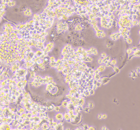
After 5 days of RANKL induction, classic multinucleated osteoclast structures can be observed under the microscope.
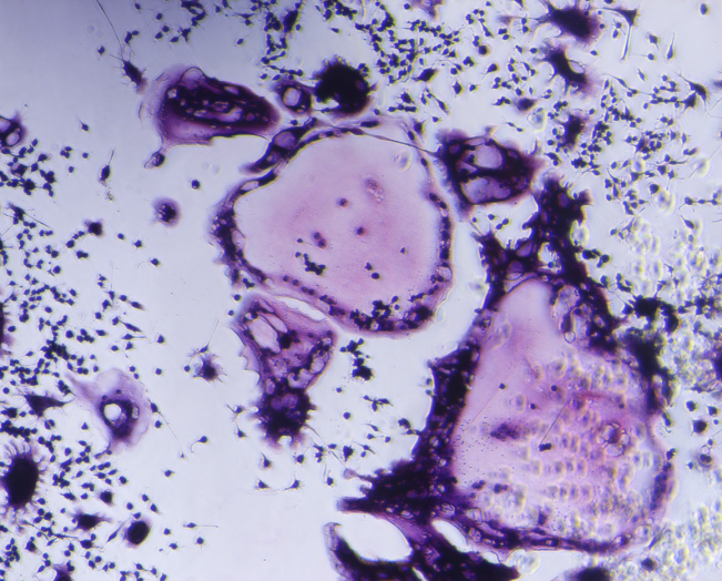
After 5 days of RANKL induction, perform TRAP staining to specifically identify osteoclasts. After staining, observe under an optical microscope. Mature osteoclasts will show red or purple-red cytoplasm.
IV. Primary Bone Marrow Cell (BMMs) Osteoclast Induction Protocol (UA090055)
Procedure:
1. Bone Marrow Cell Seeding
1.1 After lysing red blood cells from freshly harvested mouse bone marrow cells, resuspend in α-MEM complete medium containing 10% FBS and perform cell counting.
1.2 Dilute the cell density to 2 × 10⁵ cells/mL, add M-CSF to a final concentration of 30 ng/mL, and seed the cells in a 24-well cell culture plate pre-placed with sterile coverslips, adding 1 mL of cell suspension per well.
1.3 Place the plate in a 37°C, 5% CO₂ cell incubator and culture for 5 days, replacing the medium (containing 30 ng/mL M-CSF) every 2 days.
2. Osteoclast Induction and Differentiation
2.1 Aspirate the old medium. Add complete medium containing 30 ng/mL M-CSF to the negative control wells, and add complete medium containing 30 ng/mL M-CSF and 50 ng/mL RANKL protein to the positive wells.
2.2 Thereafter, replace the medium every 2 days. Add complete medium containing 30 ng/mL M-CSF to the negative control wells, and add complete medium containing 30 ng/mL M-CSF and 50 ng/mL RANKL protein to the positive wells.
3. Cell Morphology Observation and Identification
3.1 Morphology Observation: Regularly observe cell morphological changes under an inverted microscope from the start of induction. Typically, after 4 days of RANKL induction, osteoclast-like structures can be observed.
3.2 TRAP Staining Verification: After 5-7 days of RANKL induction, perform TRAP staining to specifically identify osteoclasts. Follow the TRAP staining kit instructions strictly.
After staining, observe under an optical microscope. Mature osteoclasts will show red or purple-red cytoplasm.
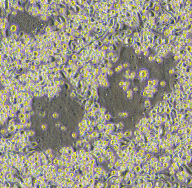
After 6 days of RANKL induction, osteoclasts under the microscope.
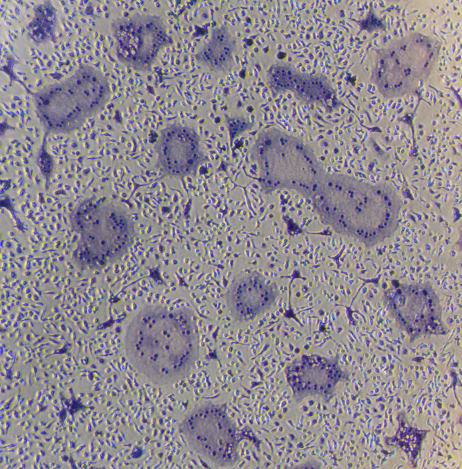
After 6 days of RANKL induction, perform TRAP staining to specifically identify osteoclasts. After staining, observe under an optical microscope. Mature osteoclasts will show red or purple-red cytoplasm.
Key Control Points for Successful Experiments
To achieve ideal induction results, in addition to strictly following the instructions, attention must be paid to the following critical details:
Cell state is the foundation. For Raw264.7 cells, use cells with low passage numbers, in the logarithmic growth phase, and in good condition, avoiding over-digestion during passaging.
For BMMs, it is crucial to aseptically, quickly, and efficiently isolate bone marrow cells from mouse femurs and tibias, and thoroughly lyse red blood cells to minimize interference.
Quality control of reagents and operations. When dissolving recombinant cytokine lyophilized powder, use sterile ultrapure water and mix gently by pipetting, avoiding repeated freeze-thaw cycles. Aliquot storage is recommended.
During the induction process, medium replacement should be performed gently to avoid aspirating loosely attached cells (especially early BMMs). Use verified high-quality fetal bovine serum throughout.
Gold standard for phenotype identification. Multinucleated morphology under the microscope is a preliminary judgment, while TRAP staining is an irreplaceable specific method for identifying mature osteoclasts.
After staining, mature osteoclasts should show distinct red or purple-red cytoplasm, with unstained nuclei.
V. Application Scenarios and Research Prospects
| Research Area | Recommended Model | Application Examples |
|---|---|---|
| Osteoclast Differentiation Mechanism | BMMs Model | Transcription factor and signaling pathway regulation studies |
| Anti-Osteoporosis Drug Screening | RAW264.7 Model | High-throughput screening of osteoclast differentiation inhibitors |
| Natural Product Activity Evaluation | Dual Model Validation | Anti-bone resorption activity studies of traditional Chinese medicine monomers |
| Bone Immune Microenvironment Research | BMMs Model | Effects of inflammatory cytokines on osteoclast differentiation |
| Gene Function Studies | RAW264.7 Model | Analysis of gene knockout/overexpression effects |
Technological Development Trends
With the advancement of organoid technology and microfluidics, in vitro osteoclast models are evolving toward 3D culture, multi-cell co-culture (with osteoblasts and immune cells), and high-throughput automation. The high activity and stability of UA BIOSCIENCE's cytokine kits provide reliable basic reagent support for establishing these advanced models.
This article is reviewed and published by the technical expert team of UA
Disclaimer: This article partially utilizes artificial intelligence assistance in its creation. If any content involves copyright or intellectual property issues, please let us know and we promise to verify and remove it as soon as possible.
Purchase recombinant protein, choose 
UA protein focuses on providing various protein reagents, raw materials, and services required for drug research and development, cell therapy, gene therapy, and basic scientific research, including drug target proteins, immune checkpoint proteins, cytokines, tool enzymes, customized protein expression, and full-length transmembrane protein development. Nanjing Univ is committed to providing customers with high-quality products and professional services, and building a High-tech Biological Enterprise with International Competitiveness.







 Business license (three certificates in one)

