




The cell-specific mechanisms of the dual role of interferon-γ in the tumor microenvironment


Interferon-gamma (IFN-γ), as the sole member of type II interferons, exhibits complex dual functions in tumor immune regulation.
- Recent Advances
- Product Information
I. Introduction
Interferon-gamma (IFN-γ), as the sole member of type II interferons, exhibits complex dual functions in tumor immune regulation. On one hand, IFN-γ exerts anti-tumor effects by activating various immune cell subsets; on the other hand, under specific conditions, it can also promote tumor progression. This functional duality depends on its concentration in the tumor microenvironment (TME), the type of target cells, and their activation state. A comprehensive analysis of IFN-γ signaling and functional outcomes in different cell types is crucial for optimizing immunotherapy strategies. In related mechanistic studies, the Mouse IFN-γ Kit (HICA) is widely used to detect IFN-γ expression levels under various experimental conditions, providing a quantitative tool to elucidate its dose-dependent and cell-specific effects.
II. Regulation of Lymphocyte Subsets by IFN-γ
(1) Cytotoxic T Cells
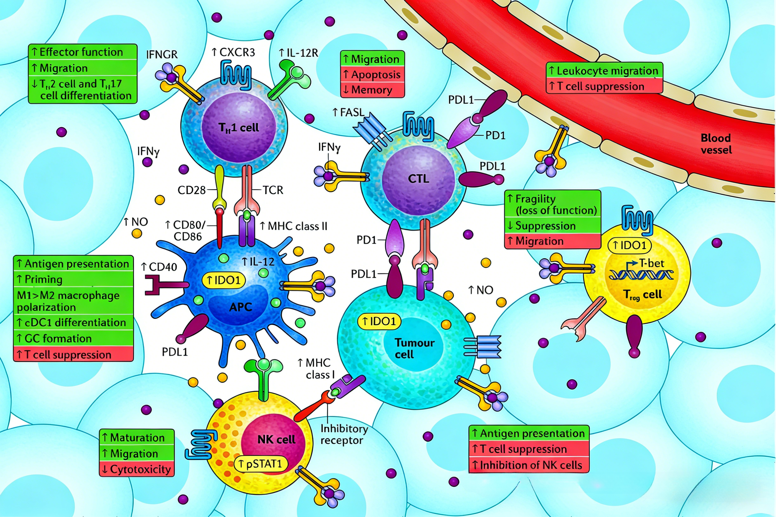
Cytotoxic T cells (CTLs) are both a major source of IFN-γ and its important target cells. IFN-γ participates in the contraction phase of CTL responses through Fas-FasL and BIM-mediated apoptosis pathways, maintaining immune homeostasis. During the CTL expansion phase, high levels of IFN-γ downregulate IL-7Rα expression via the AKT-FOXO1 pathway, limiting the formation of memory T cell pools. Although immunotherapy inducing IFN-γ production can promote the expansion of effector and memory CD8+ T cells, whether it affects long-term cell survival by regulating IFNGR and IL-7Rα expression remains to be elucidated.
In mouse tumor models, CTLs exhibit higher IFNGR expression levels than naïve T cells. IFN-γ induced by immune checkpoint inhibitor therapy can lead to activation-induced cell death, restricting the formation of effector memory cells. This mechanism may be related to tumor escape in some patients. Therefore, pre-treatment assessment of tumor burden, CTL infiltration, and IFN-γ levels can help predict therapeutic responses.
(2) CD4+ Effector T Cells
Similar to CTLs, Th1 cells producing IFN-γ downregulate IFNGR2 expression after differentiation, thereby enhancing their survival in the TME and exerting anti-tumor effects. Th1 cells inhibit polarization toward Th17 cells via T-bet-mediated suppression of RUNX1. IFN-γ suppresses Th2 polarization through dual mechanisms involving SOCS1 and T-bet, blocking IL-4 receptor signaling and GATA3 function, respectively. Under TCR stimulation, IFNGR1 and STAT1 co-localize at the immune synapse, forming a "Th1 cell-ready" state, a process inhibited by IL-4R expression in Th2 cells. Notably, PD-1 expression on tumor-infiltrating effector T cells can suppress Th1 differentiation, forming a negative feedback loop that limits IFN-γ production. Additionally, IFN-γ can promote apoptosis of effector CD4+ T cells by reducing BCL-2 expression, upregulating Fas/FasL, and inducing oxidative stress.
(3) Regulatory T Cells
In the TME, IFN-γ can drive regulatory T cells (Tregs) toward a "fragile" phenotype. These cells maintain FOXP3 expression but lose suppressive activity, thereby weakening their pro-tumor function. Treg subsets with low Nrp1 expression are associated with better prognosis in melanoma and head and neck squamous cell carcinoma patients.
III. Regulation of Innate Immune Cells by IFN-γ
(1) NK Cells
IFN-γ can activate the anti-tumor function of NK cells. Their tumor infiltration depends on IFN-γ-induced CXCR3 expression. Mice with IFNGR1 or CXCR3 knockout exhibit reduced tumor-infiltrating NK cells. IFN-γ produced by bystander T cells acts on NK cells via TRAIL, enhancing TRAIL expression through IRF1 regulation, promoting NK cell maturation and tumor-killing function.
(2) Antigen-Presenting Cells
A key anti-tumor function of IFN-γ is inducing antigen-presenting cells (APCs) to express MHC class I and II molecules, promoting tumor antigen presentation. It regulates MHC II transcription via STAT1, IRF1, and CIITA IV binding and controls MHC I expression through IRF1 and NLRC5 promoter binding. Simultaneously, IFN-γ induces the expression of co-stimulatory molecules CD80 and CD86, enhancing T cell activation.
In dendritic cells (DCs), IFN-γ drives their differentiation into cDC1 subsets, expressing CD80, CD86, MHC class I, CD40, CD54, and CCR7, and secreting IL-1β and IL-12 to promote Th1 differentiation and CD8+ T cell activation. In B cells, IFN-γ synergizes with B cell receptor and CD40 signaling to induce the germinal center transcription factor BCL-6 and collaborates with IL-12 to promote antibody class switching to IgG2a, enhancing antibody-dependent cellular cytotoxicity.
In macrophages, IFN-γ, as a classic "macrophage-activating factor," drives polarization toward the pro-inflammatory M1 phenotype. It suppresses M2 polarization by downregulating miR-3473b and induces CXCL9 and CXCL10 production, promoting immune cell infiltration and inhibiting angiogenesis.
(3) Inhibitory Effects on Myeloid Cells
Notably, IFN-γ can also induce DCs and tumor-associated macrophages (TAMs) to upregulate inhibitory molecules such as IDO and PD-L1, promoting tumor progression through metabolic regulation and angiogenesis. IDO further stimulates TGF-β production, driving Treg differentiation and proliferation. IFN-γ also induces myeloid cells to express iNOS, which breaks down L-arginine to produce nitric oxide (NO). NO exerts anti-tumor effects by inducing apoptosis but can also promote genomic instability and angiogenesis via the p53 pathway, exhibiting pro-tumor effects depending on local concentration.
IV. Direct Regulation of Tumor Cells by IFN-γ
Tumor cells are key responders to IFN-γ in the TME. Its anti-tumor effects are primarily manifested by inducing MHC class I expression and secreting chemokines CXCL9, CXCL10, and CXCL11, promoting lymphocyte migration and inhibiting angiogenesis. However, CXCL11 exhibits pro-angiogenic activity by binding to CXCR7; CXCL9 and CXCL10 promote Th1/Th17 effector function, while CXCL11 induces Th2 and Treg responses via IL-10.
Similar to APCs, tumor cells present antigens via MHC class I molecules, but MHC I can also serve as a "self" marker inhibiting NK cell killing. Immunosuppressive tumors often downregulate MHC I expression to evade immune surveillance. IFN-γ mediates pro-tumor effects by inducing PD-L1, IDO1, iNOS, Fas, and FasL expression. Tumor cells are a major source of IDO1 and NO in the TME, with iNOS expression promoting angiogenesis and FasL expression inducing apoptosis of immune effector cells. The Mouse IFN-γ Kit (HICA) is used in these mechanistic studies to quantitatively detect IFN-γ levels, providing critical data to elucidate its dose-dependent effects.
V. Which Manufacturers Provide the Mouse IFN-γ Kit (HICA)?
Nanjing UA-Bio Technology Co., Ltd. (UA-Bio) has independently developed the "Mouse IFN-γ Kit (HICA)", a high-performance in vitro detection platform specifically designed for studying key pathways in murine Th1-type immune responses and macrophage activation. This kit is engineered to accurately and efficiently quantify the immunobinding activity of murine interferon-gamma (IFN-γ) protein, offering a stable and reliable standardized solution for mechanistic research and preclinical efficacy evaluation in fields such as tumor immunology, infectious immunity, autoimmune diseases, and drug development.
| Core Product Advantages |
|---|
| High Purity and Complete Bioactivity: The kit's core components utilize high-purity, biologically active murine IFN-γ protein validated through multi-dimensional quality control. This protein maintains the correct native homodimeric conformation and full receptor-binding capacity, faithfully simulating physiologically relevant IFN-γ-mediated immune activation signals to ensure accurate, reproducible, and functionally relevant binding assay data. |
| Exceptional Batch-to-Batch Consistency and Stability: Leveraging an internationally leading recombinant protein expression platform and highly standardized purification processes, combined with stringent release quality control systems, ensures outstanding long-term stability and excellent batch-to-batch consistency. This provides robust quality assurance for long-term, continuous preclinical research and high-throughput screening. |
| Ready-to-Use Flexible Detection Platform: Based on optimized enzyme-linked immunosorbent assay (ELISA) principles, this kit provides pre-coated strips, highly specific detection antibodies, standards, and a complete set of optimized buffer systems. It features simple, rapid operation, high sensitivity, and strong specificity, making it suitable for diverse applications such as anti-mouse IFN-γ antibody/receptor antagonist screening, neutralization activity assays, competitive binding experiments, affinity analysis, and immunogenicity evaluation. |
| Comprehensive Solutions and Professional Support: We provide fully validated standard protocols, typical dose-response curves, and detailed result interpretation guidelines to help establish stable, reproducible detection workflows. Nanjing UA-Bio's expert technical team offers end-to-end professional consultation and support for research design, assay optimization, and data analysis. |
Nanjing UA-Bio Technology Co., Ltd. is committed to providing cutting-edge, high-quality core reagents and tools for immunology, cell therapy, and innovative drug development. For detailed technical specifications, validation data, or specific application inquiries regarding the "Mouse IFN-γ Kit (HICA)" (Catalog No.: UA086046), please feel free to contact us.
This article is reviewed and published by the technical expert team of UA
Disclaimer: This article partially utilizes artificial intelligence assistance in its creation. If any content involves copyright or intellectual property issues, please let us know and we promise to verify and remove it as soon as possible.
Purchase recombinant protein, choose Nanjing UA-Bio
UA protein focuses on providing various protein reagents, raw materials, and services required for drug research and development, cell therapy, gene therapy, and basic scientific research, including drug target proteins, immune checkpoint proteins, cytokines, tool enzymes, customized protein expression, and full-length transmembrane protein development. Youai is committed to providing customers with high-quality products and professional services, and building a High-tech Biological Enterprise with International Competitiveness.







 Business license (three certificates in one)