




Mechanism study of IL-6 trans-signaling mediated by hippocampal CA1 neurons in perioperative neurocognitive disorders


Perioperative neurocognitive disorders (PND) refer to newly developed cognitive impairment or worsening of pre-existing cognitive function within 12 months before and after surgery, and are one of the most common complications in elderly surgical patients.
- Recent Advances
- Product Information
I. Research Background and Scientific Questions
Perioperative neurocognitive disorders (PND) refer to newly occurring cognitive impairment or worsening of pre-existing cognitive function within 12 months before or after surgery, representing one of the most common complications in elderly surgical patients. Previous studies have shown that interleukin-6 (IL-6), as a pleiotropic cytokine with both degenerative and regenerative effects, plays a necessary and sufficient role in triggering PND in mice following aseptic trauma. However, the specific brain region localization and signal transduction type through which IL-6 exerts this effect remain unclear. This study systematically investigates the location and type of IL-6 signal transduction in a tibial fracture aseptic trauma model. During mechanistic research, the Mouse IL-6 Kit (HICA) was used to measure IL-6 and soluble IL-6 receptor (sIL-6R) levels in cerebrospinal fluid and serum samples, providing quantitative data for signal pathway analysis.
II. Two Pathways of IL-6 Signal Transduction
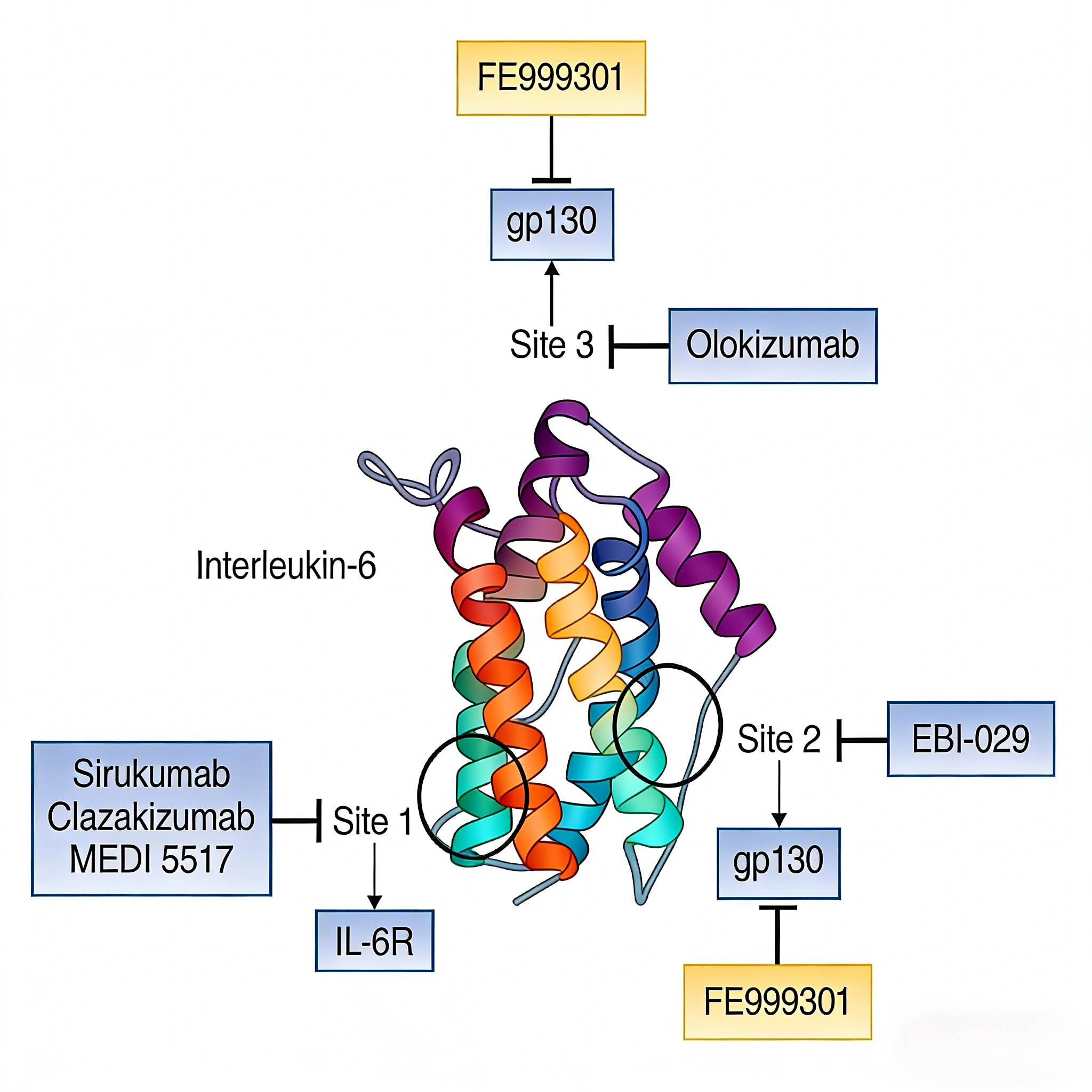
IL-6 exerts its biological effects through two distinct pathways: the classical signaling pathway involves IL-6 binding to membrane-bound IL-6 receptor α (mIL-6Rα), while the trans-signaling pathway involves IL-6 binding to soluble IL-6 receptor (sIL-6R). Both pathways require the common receptor gp130 to mediate downstream signal transduction. This study utilized wild-type and specific molecular-deficient transgenic mice, combined with a tibial fracture model under isoflurane anesthesia, to assess cognitive function through the 72-hour post-operative trace fear conditioning (TFC) test. The phosphorylation level of STAT3 (pSTAT3) in the hippocampal CA1 region at 24 hours post-operation served as an indicator of IL-6 signal activation, systematically analyzing signal pathway characteristics.
III. Association Between Hippocampal IL-6 Signaling and Surgery-Induced Memory Decline
(1) Surgery-induced cognitive impairment correlates with elevated hippocampal IL-6 levels
Wild-type mice subjected to tibial fracture intervention under general anesthesia showed a significant decrease in the percentage of freezing time during the 72-hour post-operative TFC test, indicating cognitive impairment. At 24 hours post-operation, elevated IL-6 levels were detected in hippocampal tissue, with a significant increase in sIL-6R in cerebrospinal fluid but no similar changes in peripheral blood, suggesting IL-6 signal activation within the central nervous system.
(2) Exogenous IL-6 replicates surgical effects
Lateral ventricular injection of exogenous IL-6 (400 ng) replicated the surgery-induced cognitive decline and upregulated pSTAT3 levels in the hippocampal CA1 region. This dose of IL-6 cannot cross the blood-brain barrier, confirming its central specificity. Pretreatment with IL-6R antibody BE0047 blocked the behavioral changes induced by surgery and exogenous IL-6, further validating the critical role of IL-6 signaling in PND pathogenesis.
IV. Validation of the Necessity of IL-6 Signaling in CA1 Neurons
(1) Protective effect of gp130 deletion in CA1 neurons
Both classical and trans-IL-6 signaling require the common receptor gp130. In mice with CA1 neuron-specific gp130 knockout, neither surgery nor exogenous IL-6 induced reduced freezing behavior or upregulated CA1 pSTAT3. This result clearly demonstrates that IL-6 signaling mediates PND occurrence through hippocampal CA1 neurons.
(2) Classical signaling pathway does not participate in PND induction
Experiments using systemic mIL-6Rα knockout mice revealed that surgical intervention still induced behavioral impairment and STAT3 phosphorylation in CA1 neurons. This indicates that membrane-bound IL-6Rα is not required for IL-6-mediated memory decline, and the classical signaling pathway is not involved in PND occurrence.
V. Core Role of the Trans-Signaling Pathway
(1) Exclusion analysis of sIL-6R sources
Microglia highly express IL-6Rα and are a potential source of sIL-6R. However, in microglia IL-6Rα-deficient mice, surgery still induced cognitive decline and STAT3 activation in CA1 neurons, suggesting microglia are not the primary source of PND-related sIL-6R.
(2) Protective effect of trans-signaling blockade
Application of sgp130Fc to selectively block IL-6 trans-signaling completely abolished the cognitive impairment and CA1 pSTAT3 upregulation induced by surgery and exogenous IL-6. In systemic IL-6Rα knockout mice, surgery and exogenous IL-6 also failed to induce PND phenotypes, further confirming the necessity of sIL-6R in PND pathogenesis.
VI. Feasibility Assessment of Therapeutic Interventions
(1) Impact of selective signal blockade on fracture healing
Early inflammatory responses post-fracture involve the secretion of various pro-inflammatory factors, with IL-6 playing a critical role in bone healing. The study compared the effects of non-selective IL-6 signal blockade (BE0047) versus selective trans-signaling blockade (sgp130Fc) on fracture healing. Results showed that the percentage of newly formed collagen in callus tissue was significantly higher in the sgp130Fc group than in the BE0047 group (52.2% vs. 39.7%), indicating classical IL-6 signaling is essential for fracture healing.
(2) Clinical implications of therapeutic strategies
This study reveals that IL-6 mediates PND through trans-signaling in hippocampal CA1 neurons, while classical signaling is indispensable for fracture healing. This finding suggests that selectively targeting the trans-signaling pathway may prevent PND while preserving classical signaling's bone-healing function, offering significant clinical translation potential.
VII. Which Manufacturers Provide the Mouse IL-6 Kit (HICA)?
Nanjing UA-Bio Technology Co., Ltd. (UA-Bio) has independently developed the "Mouse IL-6 Kit (HICA)", a high-performance analysis platform specifically designed for studying key pathways in murine inflammatory signal transduction and immune regulation. This kit enables precise and efficient quantitative detection of interleukin-6 (IL-6) protein immunobinding activity in mice, providing stable and reliable standardized solutions for mechanistic research and preclinical drug efficacy evaluation in autoimmune diseases, chronic inflammation, infection immunity, and related fields.
| Core Product Advantages |
|---|
| High Purity and Full Biological Activity: The kit's core components utilize high-purity, high-bioactivity murine IL-6 protein validated through multi-dimensional quality control. This protein maintains correct native conformation and full receptor complex binding capacity, accurately simulating physiological IL-6-mediated inflammatory and immune regulatory signals in mice, ensuring accurate, reproducible, and functionally relevant binding assay data. |
| Exceptional Batch-to-Batch Consistency and Stability: Leveraging an internationally advanced recombinant protein expression platform and highly standardized purification processes, combined with stringent release quality control systems, the product exhibits outstanding long-term stability and excellent batch-to-batch consistency. This provides robust quality assurance for long-term, continuous preclinical research and high-throughput screening. |
| Ready-to-Use Flexible Detection Platform: Based on optimized enzyme-linked immunosorbent assay (ELISA) principles, the kit provides pre-coated strips, highly specific detection antibodies, standards, and a complete set of optimized buffer systems. It features simple and rapid operation, high sensitivity, and strong specificity, suitable for various research applications including anti-mouse IL-6 antibody/receptor antagonist screening, neutralizing activity assays, competitive binding experiments, affinity analysis, and immunogenicity evaluation. |
| Comprehensive Solutions and Professional Support: We provide fully validated standard protocols, typical dose-response curves, and detailed result interpretation guidelines to help establish stable and reproducible detection workflows. Nanjing UA-Bio's technical team offers professional consultation and support for research design, experimental optimization, and data analysis. |
Nanjing UA-Bio Technology Co., Ltd. remains committed to providing cutting-edge, high-quality core reagents and tools for immunology, cell therapy, and innovative drug development. For detailed technical parameters, validation data, or specific application inquiries regarding the "Mouse IL-6 Kit (HICA)" (Catalog No.: UA086051), please feel free to contact us.
This article is reviewed and published by the technical expert team of UA
Disclaimer: This article partially utilizes artificial intelligence assistance in its creation. If any content involves copyright or intellectual property issues, please let us know and we promise to verify and remove it as soon as possible.
Purchase recombinant protein, choose Nanjing UA-Bio
UA protein focuses on providing various protein reagents, raw materials, and services required for drug research and development, cell therapy, gene therapy, and basic scientific research, including drug target proteins, immune checkpoint proteins, cytokines, tool enzymes, customized protein expression, and full-length transmembrane protein development. Youai is committed to providing customers with high-quality products and professional services, and building a High-tech Biological Enterprise with International Competitiveness.







 Business license (three certificates in one)